Heart
| Heart | |
|---|---|
 |
|
| System: | Both Arterial system & Venous system |
| Function: | Circulation |
| Origin: | |
| Branches: | |
| Insertion: | |
| Arterial supply: | Right and left coronary arteries |
| Venous drainage: | Coronary sinus |
| Lymphatic drainage: | |
| Innervation: | Vagus nerve |
| Vertebral levels: | |
| Search for Heart in Gray's. | |
Contents |
Basics
The pump in the circulatory system, the heart consists of specialised muscle. Gross structure has 4 chambers separated by valves. The atria fill passively from venous return, then actively expel the blood into the ventricles. The ventricles then pump the blood around the body. All cardiac myocytes have automacity, but the rate of discharge varies. The fastest in health are in the sinoatrial (SA) node, which therefore control the heart rate. This spreads a "wave" of conduction through the atria, causing contraction. The atria and ventricles are electrically isolated, except through the atrioventricular (AV) node, and occasionally accessory pathways. The AV node delays conduction into the ventricles, but then propagates it through the conducting system, down the bundle of His.
Surface markings
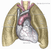
- Apex is at mid-clavicular line of 5th intercostal space.
- Inferior border passes horizontally to right sternal edge at level of sixth rib.
- Right border passes along right sternal edge between 3rd and 6th ribs.
- Left border is oblique and runs from eft sternal edge at 2nd rib to apex.
- Superior border crosses sternum between 3rd rib on right and 2nd rib on left.
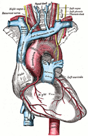
Valves
- Mitral is between left atria and ventricle
- Aortic is between left ventricle and aorta
- Tricuspid is between right atria and ventricle
- Pulmonary is between right ventricle and pulmonary artery
Heart transplant
Transplantation of the heart (or of the heart and lungs may be performed in some cases.